Ectoparasite and pathogen surveillance in Laos (TickMap 7)
 Collaborations
• U.S. Naval Medical Research Unit-2.
• Pathogen Discovery Laboratory, Institut Pasteur Paris (IPP)-France.
Funding
• U.S. Naval Medical Research Center-Asia (NMRC-A) in support of the Department of Defense Global Emerging Infections Surveillance and Response System (DoDGEIS).
• French Ministry of Higher Education and Research.
Objectives
• Survey and modern identification (ID) of Indigenous ticks and other associated arthropods, species distribution.
• Collection, ID, extraction of vector DNA for submission and development of regional repository.
• Detection of putative pathogens associated with ectoparasites from Laos.
• Building local capacities and competencies.
Background
Vector-borne diseases constitute a significant infectious disease risk for deployed military personnel and for local populations. In Laos, definitive diagnosis is often not available for vector-borne illnesses, so the infectious diseases that are a threat to military and civilian populations are not well-defined. In order to identify common and emerging vector-borne pathogens in Laos, in collaboration with NAMRU-2 Singapore (SG), we established a study to assess the distribution and infection potential of vectors (including ticks and associated arthropods). In this study, tick and associated arthropod vectors were surveyed from the environment and their associated hosts to provide biological specimens for diagnostic purposes. The samples were transported to the Institut du Pasteur (IPL) laboratory in Vientiane, where a wide range of diagnostic tests were performed to identify both the vector and the pathogens with which they were infected. In order to understand the infectious disease threats in a range of environments in Laos, IPL collected and screened specimens from 2 sites and 2 provinces throughout Laos.
Methodology
Field sites and times.
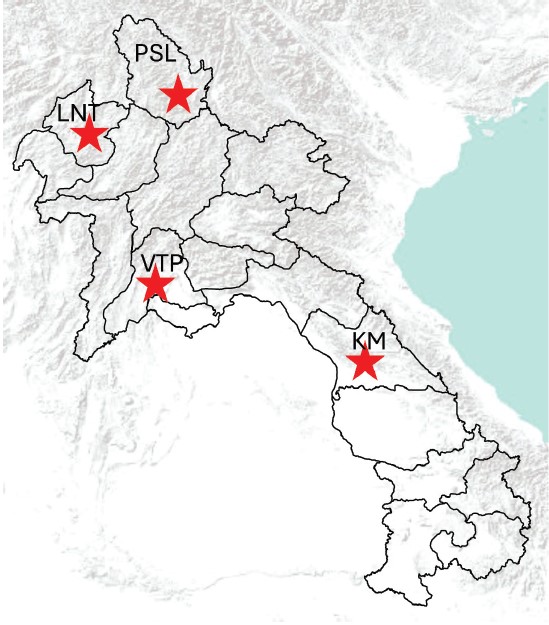
During the course of our study between 2023 and 2024, ectoparasites were collected from Khammouane, Luangnamtha, Phongsaly, and Vientiane provinces (Fig. 1).
Figure 1: Sampling sites of ectoparasites between 2023 and 2024. KM: Khammouane, LNT: Luangnamtha, PSL: Phongsaly, and VTP: Vientiane province.
Field collection procedure.
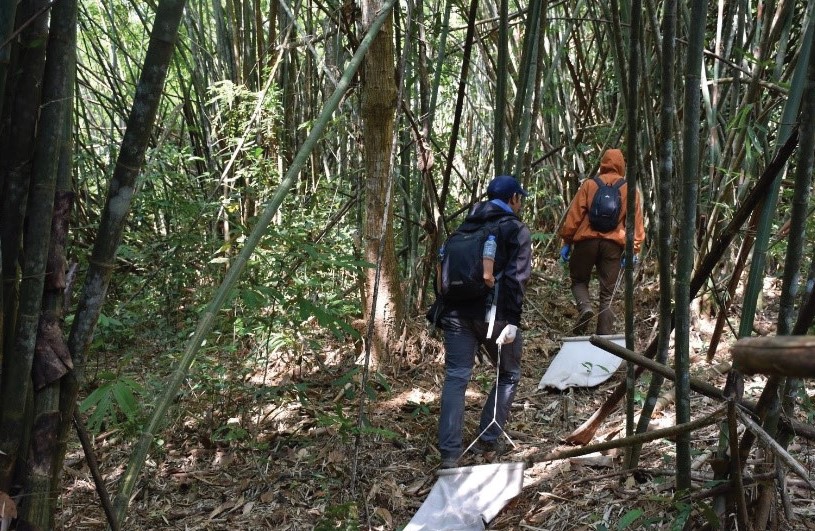
Tick dragging/flagging: Tick dragnets were swept/ dragged along the forest ground at approximately 1–2 m intervals before being examined for ticks (Fig.2). Ticks were removed from the sheets using forceps, then transferred to 1.5 ml labeled cryotubes, and stored -20°C. Our dragnet collecting was carried out in all three sites.
Figure 2: Tick dragging in forest in Phongsaly province in November 2023.
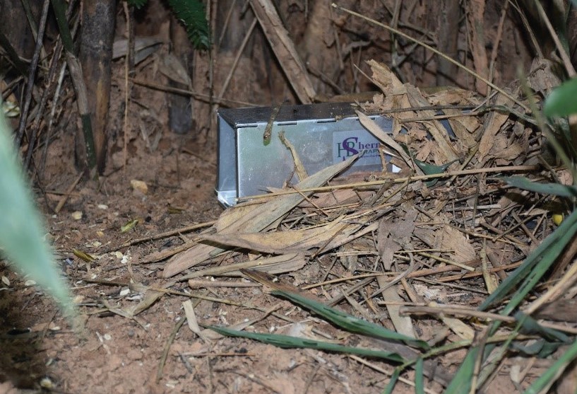
Small Mammal Trapping: In each study site, 50 Sherman traps (baited with bananas, sticky rice, or dried fish/ meat) were placed in the format of a transect according to the topography in a plantation or forest (Fig. 3).
Figure 3: Setting Sherman traps for rodent capture in forest areas.
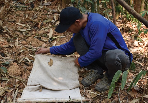
Additional tick collection was carried out by examining domestic animals (cows). The animal owners were asked to help to examine their animals. Once ticks attached to animals were found, they were collected by direct hand removal. All samples were stored at -20°C in the field and transported to the Vientiane Laboratory (IPL) using dry ice.
Laboratory work.
Sample preparation and RNA/DNA extraction.
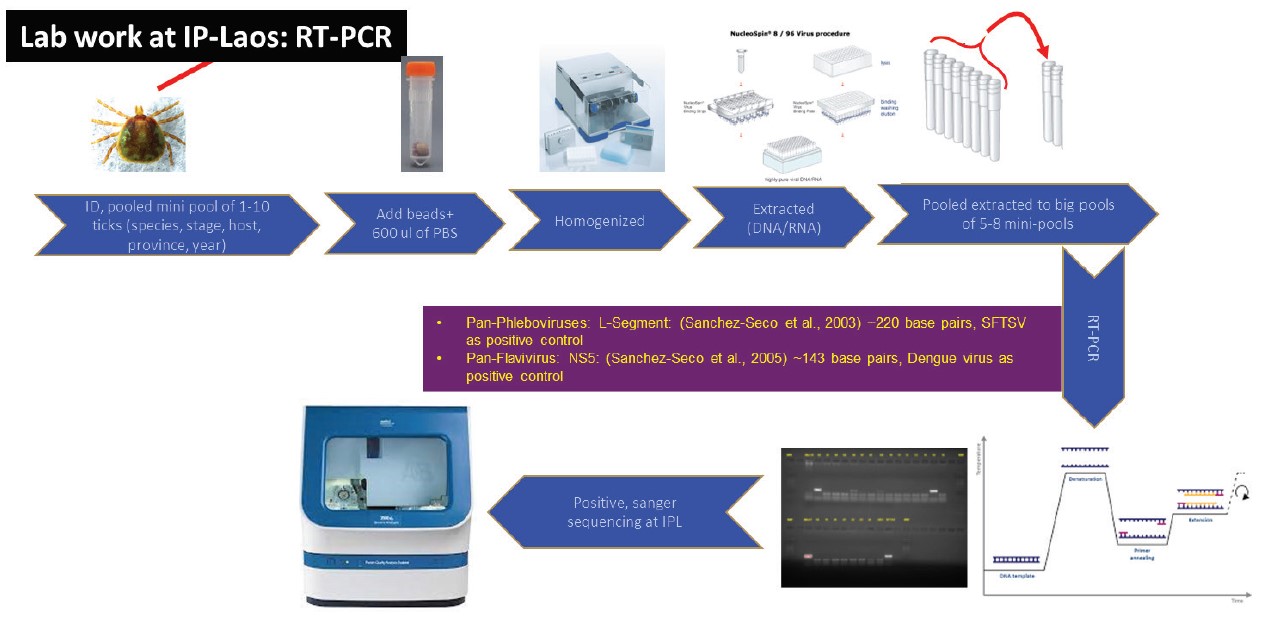
Between 1 and 10 ticks/ectoparasites were pooled together based on collection source, species, development stage, blood meal, and site. Nucleic acids from the pools were extracted as follows: specimens were placed in a 1.5 ml vial containing 1 ml of 1X cold Phosphate Buffered Saline (PBS) and Lysing Matrix A zirconium beads (MP Biomedicals). Tick pools were homogenized for 10 min at a vibration frequency of 25/s in a TissueLyser II system (Qiagen). After grinding, beads and tissues were spun down by centrifugation for 5 min at 3000 rpm. To obtain total nucleic acid (both DNA and RNA) for bacterial and viral detection by polymerase chain reaction (PCR), 100 μl of each pool was extracted and purified by using NucleoSpin® 8 Virus extraction kit following the manufacturer’s protocol. The remaining 400 μl of each pool was stored at –80°C for future studies.
Arbovirus screening at IPL.
Extracted samples were initially screened for phleboviruses and flaviviruses by RT-PCR at IPL (Fig.4) as previously described by Sanchez-Seco et al., 2003 and 2005. Samples were also tested for Jingmen tick virus (JMV) by RT-qPCR as described by Cicculli, V. et al., 2024.
Figure 4: Lab work procedure for pan-flavivirus and pan-phlebovirus screening at IP-Laos.
Implementation of rickettsia testing at IPL.
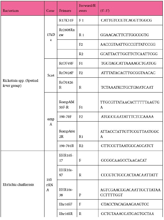
To investigate the occurrence of Rickettsia spp. from the spotted fever group in ticks, a molecular screening approach targeting the 17kDa gene (Jiang et al. 2004) was implemented at IPL. To identify Rickettsia to species level, we also examined the Sca4, and ompA genes. Ehrlichia chaffeensis and Anaplasma phagocytophilum were also included in the testing panel (Tab. 1) and Sanger sequencing was performed as previously described by Taylor et al., 2016.
Table 1: Primers used for rickettsia species identification.
Next generation Sequencing.
RNA library construction, and NGS sequencing.
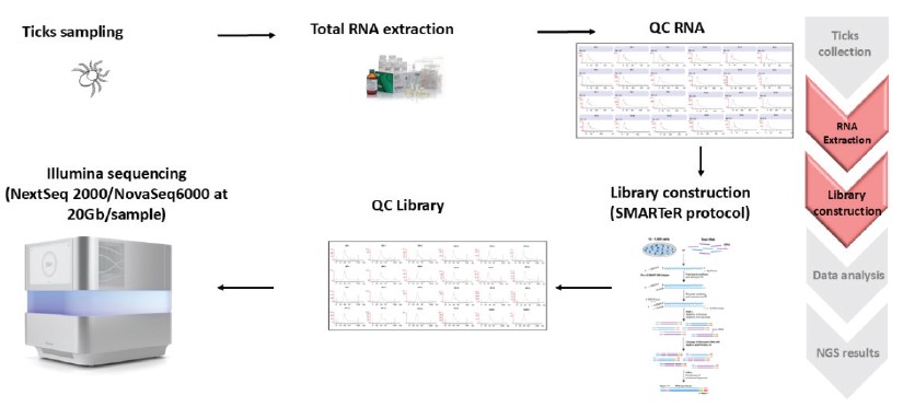
In brief, the quality and concentration of extracted samples were evaluated using Agilent TapeStation and Qubit 2.0 Fluorometer (Invitrogen, USA) respectively. Total RNA libraries were constructed using the SMARTer Stranded Total RNA-seq Kit v3-Pico input mammalian (TaKaRa). DNA library quality and concentration were evaluated using the same techniques mentioned above. Then the libraries were sent to Macrogen Asia Pacific Pte. Ltd. for NGS using platform NovaSeq6000 at 20Gb/ sample.
Figure 5: Procedure for NGS.
Bioinformatics pipeline.
The Pathogen Discovery lab (Institut Pasteur, Paris) designed a software called Microseek. It is a pipeline oriented initially for pathogen discovery but that also can efficiently detect known viruses (see https://pubmed. ncbi.nlm.nih.gov/36146797/).
Results
Species composition and abundance of ectoparasites collected between 2023 and 2024.
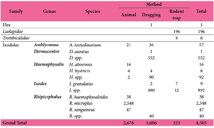
A total of 4,505 ticks and other ectoparasites were collected from two provinces, of which 2,676 were from animals, 1,606 from dragging, and 223 from rodents (see Tab. 2 below for more details). Ticks were classified into 12 species of 5 genera including Amblyomma testudinarium, Dermacentor auratus, and D. spp.; Haemaphysalis aborensis, H. hystricis, and H. spp.; Ixodes granulatus, and I. spp.; and Rhipicephalus haemaphysaloides, R. microplus, Rh. sanguineus and Rh. spp. The most abundance was Rh. microplus (See Tab. 2 below for more detail).
Table 2: Ectoparasites collected from different sources and their classification.
Pathogen screening.
Rickettsia screening at IPL.
Spotted fever group Rickettsia.
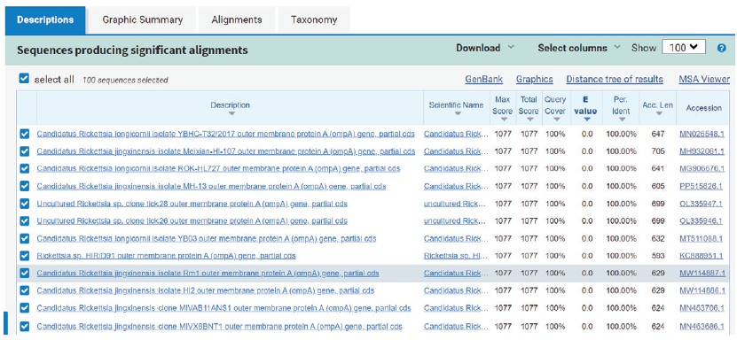
A total of 107 mini pools of ticks (corresponding to a total of 341 ticks) were screened by conventional PCR for the 17kDa gene of the spotted fever group rickettsia species. The results showed that 54 pools (145 ticks) (see Tab. 3 below) yielded the expected product size (~434 bp). So far, Sca4 and ompA genes have been successfully amplified in 15 samples of these positive tick pools. Sanger sequencing of these three genes confirmed the appartenance to the rickettsia species (see Fig.6-8 below for blast results). Sca4 gene was 100% identical to Candidatus Rickettsia Jingxinensis (Fig.7), whereas the ompA gene showed 100% identity to many Rickettsia species (Fig.8); the analysis of other additional genes such as gltA, ompB, and Fla B is needed for the precise identification at the rickettsia species level.
Table 3: Total number of tick pools and ticks screened for spotted fever group rickettsia using 17kDa gene.
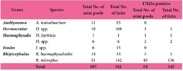
Figure 6: Blast results – 17kDa Gene from Rhipicephalus microplus.
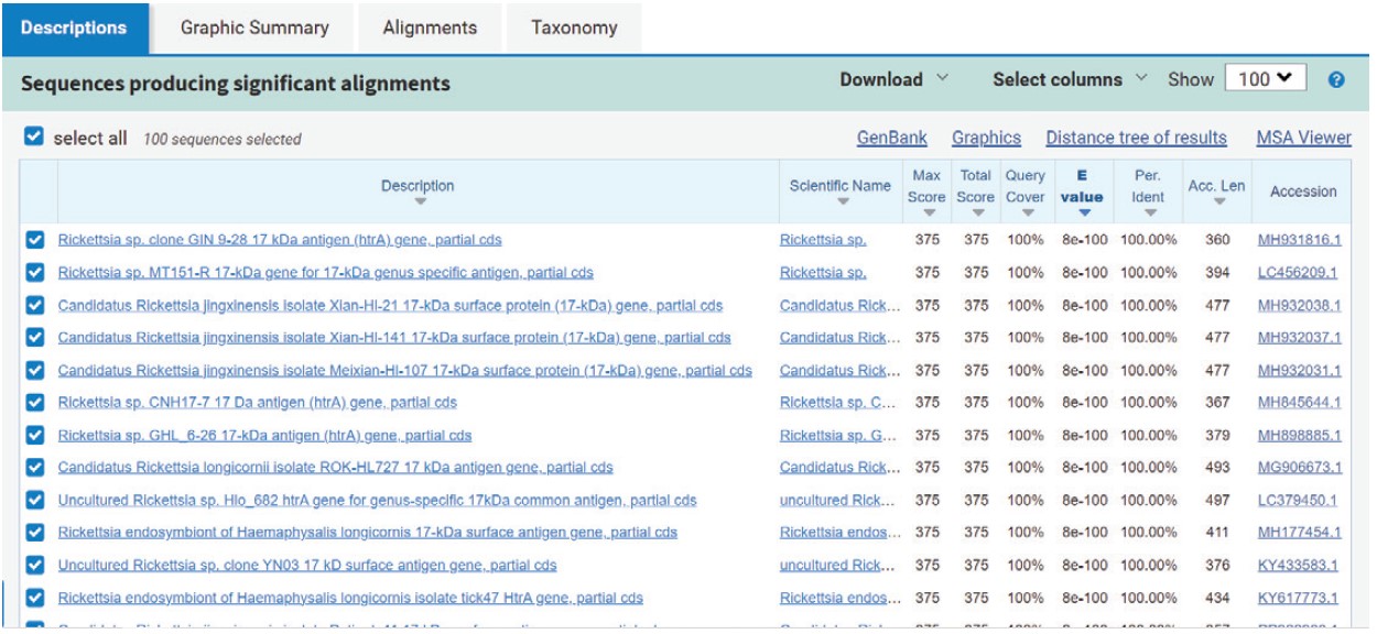
Figure 7: Blast results – Sca4 gene from Rhipicephalus microplus.
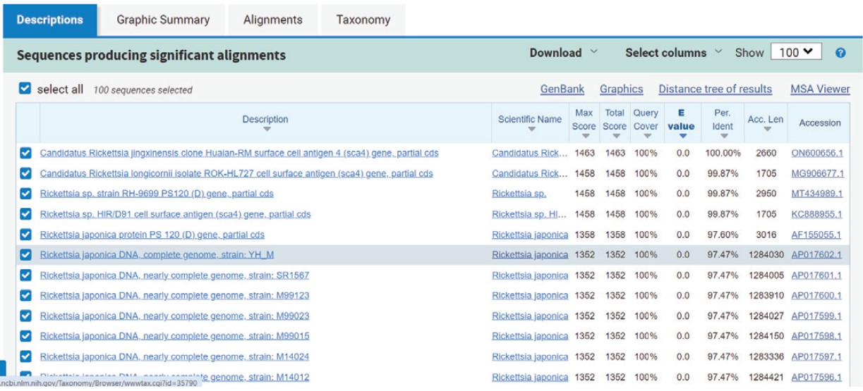
Figure 8: Blast results – ompA gene from Rhipicephalus microplus.
Ehrlichia chaff eensis.
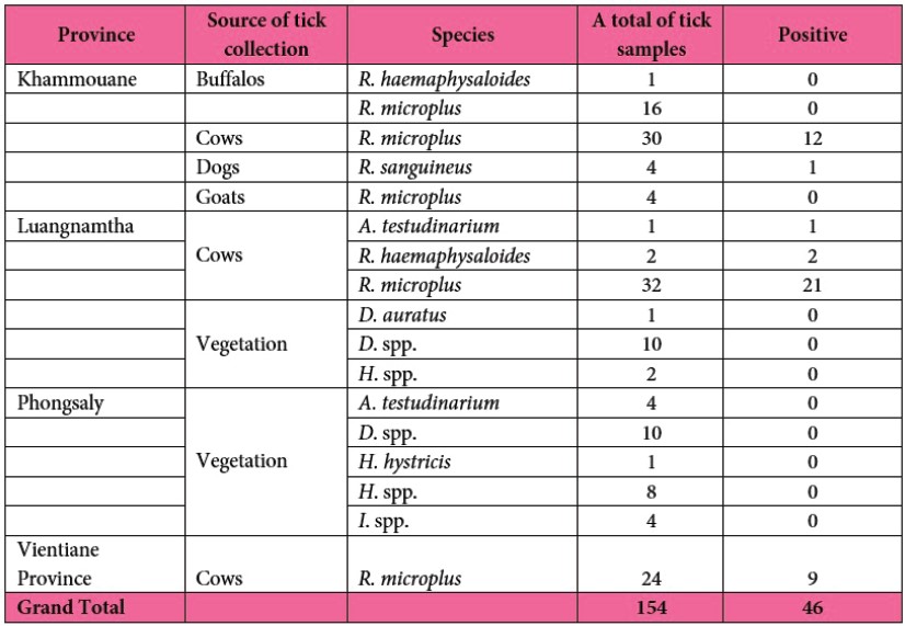
A total of 154 individual tick samples collected from various sources in four provinces of Laos were screened for Ehrlichia chaff eensis using qPCR. Of these, 46 out of 154 samples tested positive (Tab. 4).
Pan-flaviviruses and pan-phleboviruses screening by RTPCR.
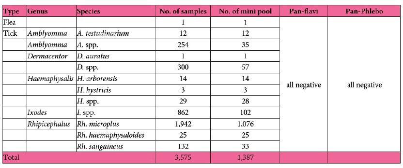
So far, a total of 1,387 mini pools of 3,575 ticks have been pooled and initially screened for pan-faviviruses and pan-phleboviruses. All samples tested negative (see Tab. 5 below).
Table 5: Total number of tick pools and ticks screened for pan-flaviviruses and pan-phleboviruses.
Jingmen tick virus detection among ticks in Laos.
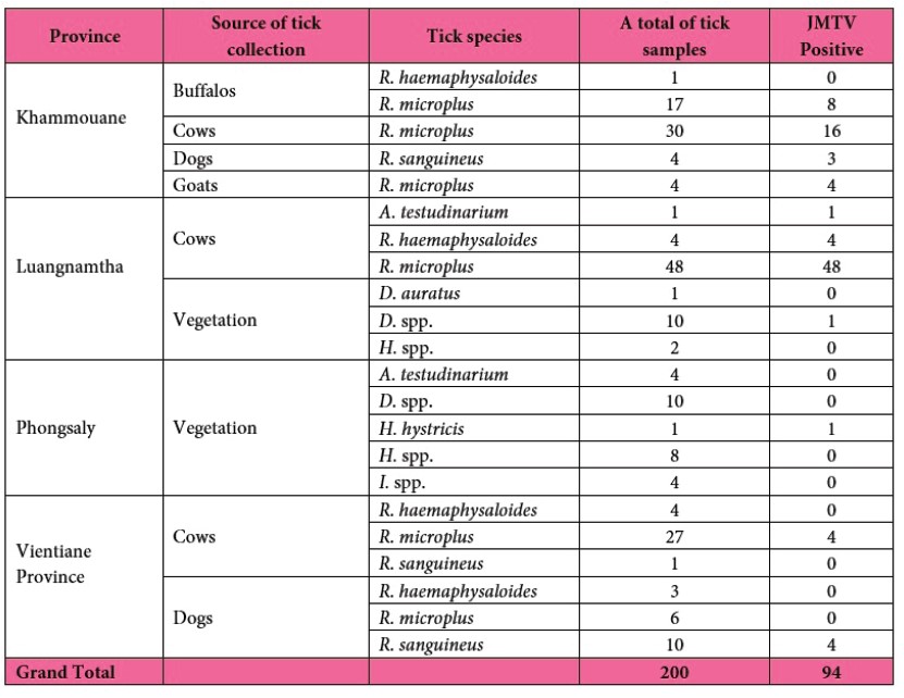
A total of 200 individual tick samples collected from different sources in four provinces of Laos were screened for JMTV. Among them, 94/200 were positive a with high prevalence in Rhipicephalus ticks collected from domestic animals (Tab. 6).
Table 6: Total number of ticks screened for Jingmen tick virus.
Tick virome analysis by metagenomic approaches.
Preliminary analysis of data generated by NGS from 2019 to 2023.
From 2019 to 2023, despite limited funding allocated for NGS, a total of 94 big pools comprising 3,424 ticks were collected from 5 provinces including Borlikhamxay, Luangnamtha, Oudomxay, Phongsaly and Xiengkhouang (Fig.9) were submitted to NGS. Eight pools containing mixed tick genera or collection sources were excluded from the analysis (red color in Tab.7).
Virome composition and diversity (viral families).
Data from the remaining 86 tick pools revealed virusrelated sequences assigned to at least 62 known virus families (heatmap of top 50 viral family shows in Fig.9), including at least six families of particular interest: Flaviviridae, Rhabdoviridae, Phenuiviridae, Peribunyaviridae, Orthomyxoviridae and Nairoviridae. The most abundant viral family was Flaviviridae (Fig.10).
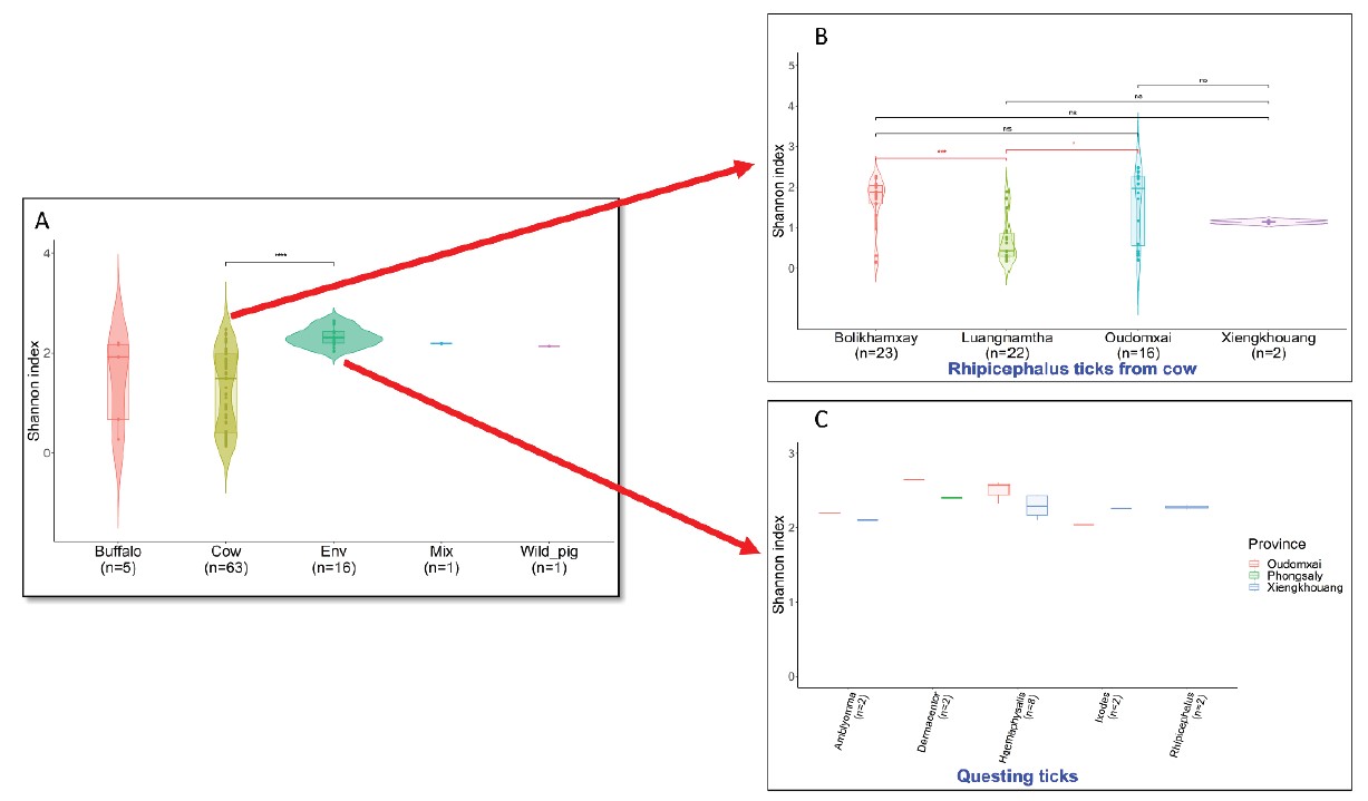
The Shannon diversity index varied based on collection sites and hosts. For Rhipicephalus ticks from cows, virome diversity was significantly lower in Luangnamtha compared to other provinces. No significant differences in diversity were observed for ticks collected from vegetation across sites or genera (see Fig.12). Genetic characterization of the viral families of interest and more detailed analysis are ongoing.
Figure 9: Map of tick samples submitted to NGS analysis between 2019 and 2023.
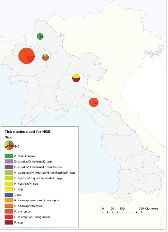
Table 7: Tick samples submitted to NGS analysis between 2019 and 2023. Pools with mixed tick genera or collection sources were removed from the analysis (red color).
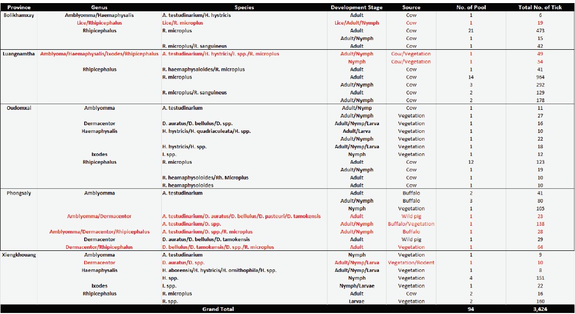
Figure 10: Heatmap showing the top 50 virus families detected in 86 tick pools.
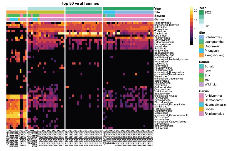
Figure 11: Proportion of virus families detected from 86 tick pools. Red stars indicate families of interest.
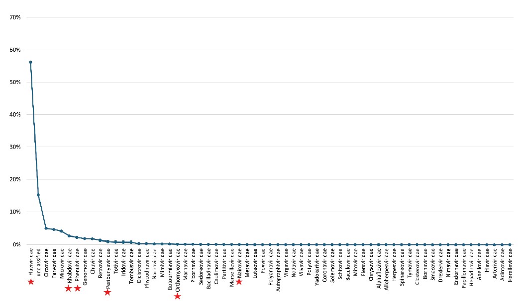
Figure 12: Shannon diversity indexes of viral families among ticks collected from different sources (A). Shannon diversity indexes among Rhipicephalus ticks from cows by provinces (B). Shannon diversity indexes among ticks collected by dragging by genus and sites (C).
To continue the surveillance of ticks and tick-borne pathogens in Laos and investigate the influence of ecological factors on pathogen circulation and cocirculation, NGS analysis at the individual tick level is planned. So far, 60 tick samples have been selected for library preparation.
Conclusion and perspectives
This study provides comprehensive data on tick and ectoparasite diversity and their potential as vectors of pathogens in Laos. Between 2023 and 2024, 4,505 ticks and ectoparasites were collected and identified, revealing 12 species across five genera. Notably, Rhipicephalus microplus was the most abundant species. Molecular screening found the presence of Candidatus Rickettsia jingxinensis in Rhipicephalus ticks, alongside preliminary detection of Ehrlichia chaffeensis. However, no Panflavivirus or Pan-phlebovirus sequences were detected. NGS analysis of tick samples collected from 2019 to 2023 identified sequences from 62 virus families, including families of interest such as Flaviviridae, Phenuiviridae, and Nairoviridae, highlighting tick virome diversity across provinces and hosts in Laos.
Future research will prioritize the genetic characterization of viruses identified through NGS, focusing on viral families of medical and veterinary importance. In 2024-2025, individual-level tick NGS strategies will be employed to improve the pathogen detection and the understanding of the ecological factors driving the circulation of tick-borne pathogens.
A key focus will be also the development and optimization of molecular techniques for the detection of known human pathogens such as Rickettsia, and Ehrlichia chaffeensis. Improved protocols for amplifying other genes such as gltA, ompB, flaB of spotted fever group Rickettsia species and more sensitive primer sets for E. chaffeensis will be essential for accurate identification and confirmation.
These methodological improvements will support a deeper understanding of the circulation of tick-borne pathogens, aiding in effective surveillance and control efforts for these diseases in Laos.
In next coming years, we will also continue to study the climate change impacts on ticks and tick-related pathogens in Lao PDR in order to model the transmission dynamics of tick-borne diseases and support the development of a country-wide and/or regional surveillance system to prevent the spread of these vector-borne diseases in the human population, especially in the poor and rural areas that are more at risk.
References
Jiang J., Chan T., Temenek J.J., Dasch G.A., Ching W. and Richards A.L. (2004). Development of a Quantitative Real-time Polymerase Chain Reaction Assay Specific for Orentia Tsutsugamushi. American Journal of Tropical Medicine and Hygiene, 70(4): 351-356.
Nadchatram, M. and A. L. Dohany. 1974. A pictorial key to the subfamilies, genera and subgenera of Southeast Asian chiggers (Acari, Prostigmata, Trombiculidae). Bulletin from the Institute for Medical Research Federation of Malaysia, 16: 1–67.
Nuttall, G. H. F., W. F. Cooper, C. Warburton, L. E. Robinson, and D. R. Arthur. 1926. Ticks: pt. IV. The genus Amblyomma. Cambridge University Press. Sanchez-Seco, M.P., et al., Detection and identification of Toscana and other phleboviruses by RT-nested-PCR assays with degenerated primers. J Med Virol, 2003. 71(1): p. 140-9.
Tanskull, P. and I. Inlao. 1989. Keys to the adult ticks of Haemaphysalis Koch, 1844, in Thailand with notes on changes in taxonomy (Acari: Ixodoidea: Ixodidae). J Med Entomol 26(6): 573–600.
Yamaguti N., V. J. Tipton, H. L. Keegan, and S. Toshioka (1971). Ticks of Japan, Korea, and the Ryukyu islands. Brigham Young University Science Bulletin, Biological Series 15(1):1.