Assessment of the potential vector threat of bat-borne pathogens and the host-associated ectoparasites in the provinces of Vientiane and Khammouane of the Lao PDR (Bat Map project)
Funded by the U.S. Naval Medical Research Center-Asia (NMRC-A) in support of the Department of Defense Global Emerging Infections Surveillance and Response System (DoD-GEIS)
Project coordinator:
+ Dr. Paul Brey, Director, Institut Pasteur du Laos, Vientiane, Lao PDR
+ Dr. Ian Sutherland, Chief of Entomological Sciences, U.S. Naval Medical Research Center – Asia, Sembawang, Singapore
+ Dr. Jeffrey Hertz, Chief of Entomological Sciences, U.S. Naval Medical Research Center – Asia, Sembawang, Singapore
Member of staff:
+ Khamsing Vongphayloth, Research entomologist, Institut Pasteur du Laos
+ Khaithong Lakeomany, Technician entomologist, Institut Pasteur du Laos
+ Nothasine Phommavanh, Technician entomologist, Institut Pasteur du Laos
Collaborators:
+ Marc Eloit , Virologist, Pathogen Discovery Unit, Institut Pasteur Paris
+ Paul Newton and Matthew Robinson, LOWMRU Wellcome Trust Vientiane Lao PDR
+ Marc Grandadam, Arbovirology Lab, Institut Pasteur du Laos
Background
The biological and social attributes of bats make them ideal reservoirs for emerging infectious diseases (EIDs). Bats represent the largest number within mammalian species (>925), occupy a variety of ecological niches, roost in large numbers, and are able to disperse over wide geographical distances. Until now, no reference data has been available regarding the viral and pathogen make-up of bats, their ectoparasitic fauna, and their potential to be an EID source within Laos.
Vector-borne diseases, including bat-borne agents, constitute a significant infectious disease risk for local populations. In Laos, definitive diagnosis is often not available for vector-borne illnesses, thus infectious diseases that are a threat to populations remain misdiagnosed and unreported. In order to identify common and emerging vector-borne pathogens in Laos, NMRC-A Singapore (SG) has established a study to assess the distribution and infection potential of bat ectoparasites and bat-borne pathogens. In this study, bat ectoparasites and associated arthropod vectors were surveyed to provide biological specimens for diagnostic purposes. The samples were transported to the Institut Pasteur du Laos (IPL) laboratory in Vientiane, where a wide range of diagnostic tests were performed to identify both the vector and the pathogens with which they may be infected. This study is known as the “Vector mapping of bat-borne ectoparasites and pathogens in the Lao PDR” (BatMap).
Khammouane and Vientiane provinces were uniquely positioned to serve as a study sites for BatMap and include a large protected area bordering Vietnam in south-central Laos, a region in which many causes of vector-borne and other illnesses are not diagnosed, and from which new infectious diseases may emerge.
In this report, we updated our activities of the project including field collection, identification, extraction, and development of molecular products for Lao repository, and reference for future studies.
Objectives
Obj 1: help address the knowledge gap in bat-borne virus and ectoparasite biology, identification, and distribution in Vientiane and Khammouane provinces.
Obj 2: conduct molecular biology detection studies for a panel of viruses and bacteria in collected specimens.
Obj 3: provide / improve infectious disease surveillance competencies (building of local capacity and competencies).
Methods
Field site collection
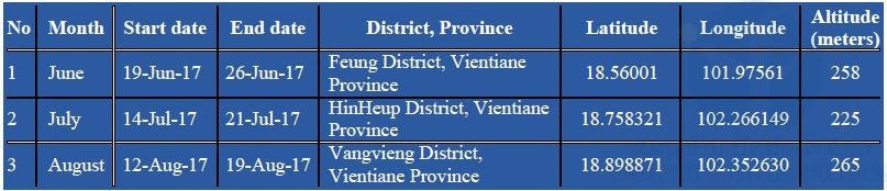
Due to administrative/authorization delays, our first field collection of bats was carried out late, in June (Q4 of the project). In collaboration with the Faculty of Environmental Sciences, National University of Laos, three field missions have been completed in three districts of Vientiane Province.
Figure 1. Map of field sites (red area) in Vientiane Province.
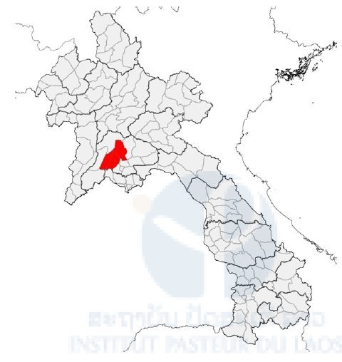
Table 1: Field site collection
Animal capture for research statement
The bat study was approved by the Institutional Animal Care and Use Committee (IACUC) (Protocol number: EXM-17-15-NAMRU-2). For the Lao side, the authorization agreement for carrying out bat capture was approved by the wildlife authorities of the Department of Forest Resource Management (DFRM), and The Ministry of Agriculture and Forestry Lao PDR, No.0174/DFRM, issued on January 2017.
Bat collection and identification procedure
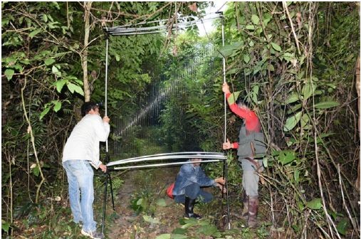
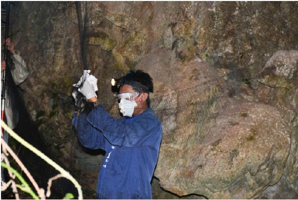
Bats were collected using four-banks harp traps (Francis, 1989), mist nets and hand (hoop) net. Harp traps were set across natural trails and over small streams in the forest understorey, and at the entrance of caves in relatively concealed conditions. Mist nets were set at the similar place to harp traps, but were more often used in opened space.
Both harp traps and mist nets were set before sunset and were left over night, except mist nets which were closed around 22.00 hour.
Captured bats were held individually in cloth bags. Species, sex, age (adult or juvenile) and reproductive condition (pregnant or lactating) were determined in the field. Species were identified following Francis (2008), Csorba et al. (2003), and Corbet and Hill (1992). Adults or juveniles were identified by the presence of unfused epiphyses of the phalanges and metacarpal joints (Brunet-Rossinni and Wilkinson, 2009). The reproductive status of female bats was determined by examining the nipples (Racey, 2009). Some external characters, including forearm length (FA), were measured using caliper and body mass (W) were taken using a Pesola spring balance. Most bats were marked with wing bands for individual identification and were released at the captured point within 12 hours.
For each species, two or three specimens were collected to confirm identification. Bats were euthanized using chloroform. Specimens were fixed in 95% ethanol in the field and were transferred to 70% ethanol when they are brought back to the laboratory. All of specimens were registered and catalogued in the Zoological Collection of the Faculty of Environmental Sciences, National University of Laos, Lao PDR.
Bat sampling procedure
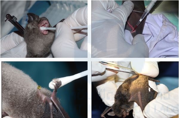
Live bats were placed into porous cotton bags (with draw-string mouths) and kept in a cool dry place until sampling time, not to exceed 6 hours. The following basic set of samples were collected from each bat: (i) Saliva (oropharyngeal swab): 1 aliquot; (ii) Feces (fresh fecal sample or rectal swab): 1 aliquot; (iii) Blood: 1 aliquot; (iv) Urine (urogenital swab): 1 aliquot ; and (v) Ectoparasites : 1 aliquot. The whole blood (serum; rbc/wbc pellet) was diluted 1/10 of PBS 1x, then centrifuged by mini centrifuge machine and then divided in two aliquots (serum and blood cells). Ectoparasites were collected using flame-sterilized forceps. All samples were stored at – 20°C then transported to IP-Laos in Vientiane and stored at – 80°C until laboratory analysis.
Lab activity
This study focused on some genus of Arbovirus and Non-arbovirus which was commonly found in bats from previous studies such as: Flavivirus, Alphavirus, Phlebovirus, and Filovirus by using appropriate techniques to detect each viral genus.
Extraction
Total nucleic acids (DNA/RNA) were extracted from bio-samples (plasma, saliva, urine, anal swab) of bats by using Nuclo Spin®8 Virus (MACHEREY-NAGAL, Germany) in extraction room. Before the assay, tube of saliva, urine and anal swabs were added 400μL of 1x PBS (phosphate buffer saline), then 150 μL were used for the nucleic acids the extraction.
Lab technique and procedure assay of Arbovirus genus
Multiple sets of primers have been selected and tested for screening specimens for the presence of phlebovirus, flavivirus, and alphavirus sequences by means of conventional nested RT-nPCR as previously described (Sanchez-Seco, Rosario et al. 2001; Sanchez-Seco, Rosario et al. 2005). Amplicons were analyzed by agarose gel electrophoresis. Positive controls were included in all series using RNA extracted from culture supernatants of reference strains (i) for Alphavirus: Sindbis, Chikungunya and Me Tri, (ii) Phlebovirus: strain IPL-13-214 and (iii) Flavivirus dengue and Japanese encephalitis.
Results
Bat capture
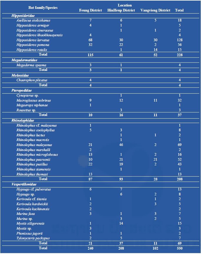
A total of 550 bats were captured during our three missions from three districts in Vientiane Province. Bats were classified into 35 species (Table 2), of which 26 species (240 bats) were collected from Feung District, 25 species (208 bats) were collected from HinHeup District, and 16 species (102 bats) were collected from Vangvieng District.
Table 2: Total number of bat captures collected during three field missions in three districts, Vientiane Province,.
Bat sample sampling
Bat biological samples
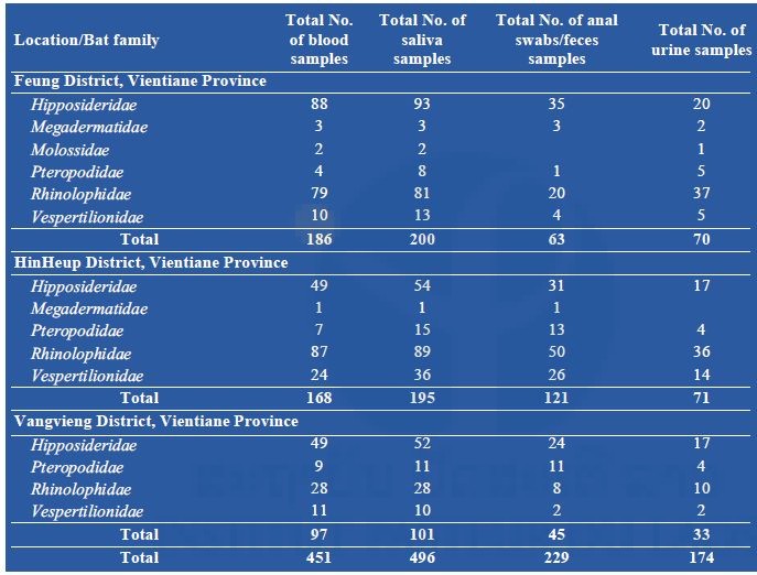
A total of 496 saliva, 451 blood, 229 anal swabs/feces, and 174 urine samples were taken from 498 bats belonging to 6 families: Hipposideridae, Megadermatidae, Molossidae, Pteropodidae, Rhinolophidae, and Vespertilionidae (Table 3).
Table 3: Total number of bat samples (saliva, blood, anal swabs/feces, and urine) collected during three field missions in three districts, Vientiane Province.
Bat ectoparasites
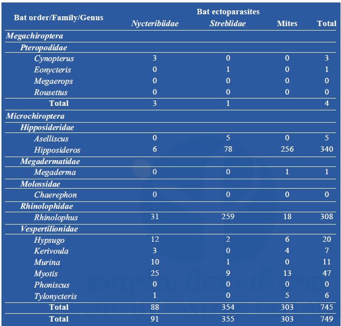
From 550 bats captured and examined for ectoparasites (Table 2 above), a total of 749 ectoparasites were collected, divided between bat mites (303) and two families of batflies: Nycteribiidae (91) and Streblidae (355) (Table 4 below).
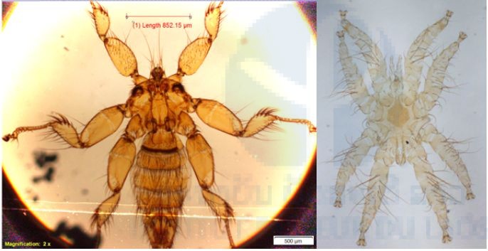
Table 4: Total number of ectoparasites (batflies and mites) collected during three field missions in three districts, Vientiane Province.
Arboviral screening
So far, a total of 186 plasma samples [plasma obtained from whole blood (serum; rbc/wbc pellet) diluted 1/10 of PBS 1x], collected from Feung District, have been submitted to total nucleic acid extraction. Of 186 samples belonging to 6 families—Hipposideridae, Megadermatidae, Molossidae, Pteropodidae, Rhinolophidae, and Vespertilionnidae—tested by RT-nested PCR techniques for the presence of arbovirus sequences (pan flavi, pan alpha, and pan phlebo), Alpha and Flavivirus sequences were not detected in our first series. Phlebovirus RNA was detected in three bats (1.61%), Rhinolophus pearsonii (n = 2/9), and Hipposideros larvatus (n = 1/56).
Discussion and ongoing activities
Apart from the administrative delays, our project continues to move forward with no currently observed problems or major obstacles. We have successfully completed our field collection from three sites in Vientiane Province with 451 blood, 496 saliva, 229 anal swabs/feces, and 174 urine samples from 550 bats captured. A total of 749 ectoparasites from 550 bats were also collected. Because of these administrative delays, it may not be possible to carry out the fourth field mission in Khammouan Province. So far, a total of 186 plasma samples from the first field collection have been extracted and screened for arboviruses; the remaining samples are ongoing. Bat samples will also be screened for the presence of other non-arboviruses, Filovirus including Ebola.
We will continue our laboratory work to identify those ectoparasites in to genus and species if possible, especially for the batflies, and submit those samples for DNA/RNA extraction. Once bat and ectoparasite samples have been extracted, half of each sample were transferred to the LOMWRU laboratory for bacterial (Bartonella sp.) investigations.
Our work needs a six-month no-cost extension because of the delay in our field collection approvals.
References
Brunet-Rossinni, A.K. and G.S. Wilkinson. 2009. Methods for age estimation and the study of senescence in bats. Pp: 315-325, in Ecological and behavioural methods for the study of bats, 2nd edition (T.H. Kunz and S. Parsons, eds). The Johns Hopkins University Press, Baltimore, xvii + 901 pp.
Corbet, G.B. and J.E. Hill. 1992. The mammals of the Indomalayan region: a systematic review. Natural History Museum Publications and Oxford University Press, 488 pp. Csorba, G., P. Ujhelyi, and N. Thomas. 2003. Horseshoe bats of the World (Chiroptera: Rhinolophidae). Alana Books, xxxii + 160 pp.
Francis, C.M. 1989. A comparison of mist nets and two designs of harp traps for capturing bats. Journal of Mammalogy, 70: 865-870.
Francis, C.M. 2008. A field guide to the mammals of Thailand and South-east Asia. New Holland Publishers (UK) Ltd and Asia Books Co., Ltd, Bangkok, Thailand, 392 pp.
Racey, P.A. 2009. Reproductive assessment of bats. Pp: 249-264, in Ecological and behavioural methods for the study of bats, 2nd edition (T.H. Kunz and S. Parsons, eds). The Johns Hopkins University Press, Baltimore, xvii + 901 pp.
Sanchez-Seco, M. P., D. Rosario, C. Domingo, L. Hernandez, K. Valdes, M. G. Guzman, and A. Tenorio. 2005. Generic RT-nested-PCR for detection of flaviviruses using degenerated primers and internal control followed by sequencing for specific identification. J Virol Methods 126(1 -2): 101-109.
Sanchez-Seco, M. P., D. Rosario, E. Quiroz, G. Guzman and A. Tenorio. 2001. A generic nested-RT-PCR followed by sequencing for detection and identification of members of the alphavirus genus. J Virol Methods 95(1-2): 153-161.
Sanchez-Seco, M. P., J. M. Echevarria, L. Hernandez, D. Estevez, J. M. Navarro-Mari, and A. Tenorio. 2003. Detection and identification of Toscana and other phleboviruses by RT-nested-PCR assays with degenerated primers. J Med Virol 71:140-149.
References
Brunet-Rossinni, A.K. and G.S. Wilkinson. 2009. Methods for age estimation and the study of senescence in bats. Pp: 315-325, in Ecological and behavioural methods for the study of bats, 2nd edition (T.H. Kunz and S. Parsons, eds). The Johns Hopkins University Press, Baltimore, xvii + 901 pp.
Corbet, G.B. and J.E. Hill. 1992. The mammals of the Indomalayan region: a systematic review. Natural History Museum Publications and Oxford University Press, 488 pp. Csorba, G., P. Ujhelyi, and N. Thomas. 2003. Horseshoe bats of the World (Chiroptera: Rhinolophidae). Alana Books, xxxii + 160 pp.
Francis, C.M. 1989. A comparison of mist nets and two designs of harp traps for capturing bats. Journal of Mammalogy, 70: 865-870.
Francis, C.M. 2008. A field guide to the mammals of Thailand and South-east Asia. New Holland Publishers (UK) Ltd and Asia Books Co., Ltd, Bangkok, Thailand, 392 pp.
Racey, P.A. 2009. Reproductive assessment of bats. Pp: 249-264, in Ecological and behavioural methods for the study of bats, 2nd edition (T.H. Kunz and S. Parsons, eds). The Johns Hopkins University Press, Baltimore, xvii + 901 pp.